Diabetes resources
Contact us to book these resources for your event or training session.
|
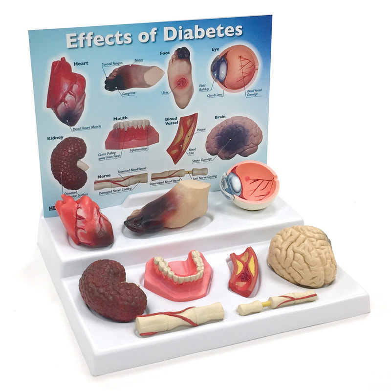
Effects of diabetes [model]
Raise awareness of the importance of proper diabetes management. This informative display features sculpted replicas of a heart, kidney, brain, eye, lower set of teeth, foot, artery, and nerves. The accompanying key card includes detailed information on the back about how diabetes damages each organ. Perfect for health fairs, doctors’ surgeries, classrooms, and diabetes educators. |

|
Title: Diabetes model 4 piece set- disease related vascular effects [model]
Description: Four piece hinged model illustrates the vascular effects of diabetes on the kidneys, arteries, nerves and eyes, ideal for patient education. |

|
Title: A1C Levels
Use this essential model to explain A1C levels to patients. One half of the model represents healthy blood glucose and A1C levels by depicting a small number of glucose particles and a red blood cell model that has few glucose particles attached to it. In contrast, the model’s other red blood cell contains excess glucose particles and floats through simulated plasma full of glucose particles, representing high A1C and blood glucose levels. |
|
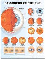
Title: Disorders of the eye anatomical chart [laminated poster]
Description: A large illustration of normal eye anatomy including normal sagittal section of the eye, close-up of a normal retina and close up of a normal optic disc are shown. Detailed illustrations of the following disorders are included; blepharitis; conjunctivitis; corneal ulcers; cataract; vitreous floaters; retinal tear and detachment; macular degeneration; diabetic retinopathy; melanoma; glaucoma. It also shows esotropia and exotropia. Size 20'' x 26'' |